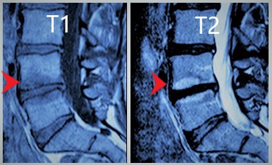
Marrow changes of the spine follow a pattern that assists the radiologist in determining the degenerative status of the lumbar spine. These marrow changes abutting the vertebral cortex are frequently seen with disk disease and are rare in asymptomatic patients. The early marrow changes are thought to be most symptomatic in up to 70% of patients and associated with mild instability.
The vertebral bone is made up of red marrow and yellow marrow in addition to cortical bone. The differences in the marrow and cortex can be identified on MRI and thus can help with the staging of the abnormality. When there is edema early on, we will see a bright signal on the fluid sequence. Over time there is diminished blood flow which causes the red marrow to convert to yellow marrow. When the process is chronic then we see sclerosis along the bone endplates which will be a dark signal on both sequences.
Although the marrow changes cannot tell the exact age of the degenerative changes, they can tell you if the process is relatively new versus old as they are related to degenerative disk disease. These marrow changes are just one of many things the expert radiologists at The Medical Consultant use to evaluate your back pain and injuries.


